Leaps forward: lessons learned from the clawed frog

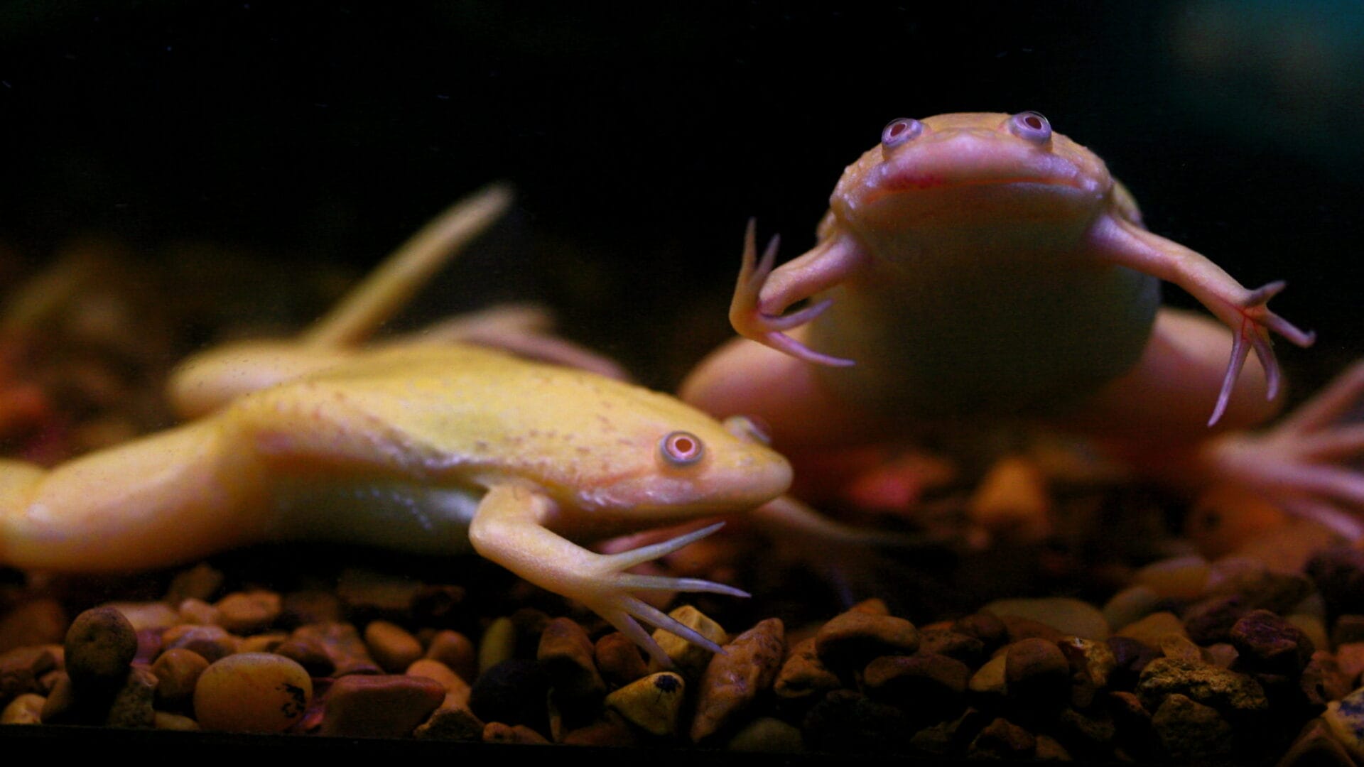
Without the African clawed frog, our understanding of early development would not have progressed to where it is today.
- The African clawed frog Xenopus laevis has many benefits as a model organism, from the ease in which they can be kept, to their abundant supply of large, robust eggs that can be simply manipulated in the lab.
- Much of our current knowledge about the early development of vertebrates comes from studies using amphibians like the clawed frog.
Key terms
Model organism
A species that has been widely studied in biology, usually because it is easy to maintain and breed in a laboratory setting and has particular experimental advantages.
Genome
The complete set of genetic instructions required to build and maintain an organism.
DNA
(deoxyribonucleic acid) A molecule that carries the genetic information necessary to build and maintain an organism.
DNA sequencing
The process of determining the order of bases in a section of DNA.
The early years of embryonic research
The history of amphibians in embryology research stretches back to the 1880s, when German embryologist Wilhelm Roux removed one cell of a two-cell frog embryo. The result was half-embryos that were unviable and had the left or right side only – indicating that the two original cells had different fates.
At this time, German scientists were undeniably the world experts in embryology – the study of development from fertilisation to embryo. During these early years, newts, salamanders, frogs and sea urchins were used to understand early development.
Embryology research before the Second World War was hampered by a lack of eggs, which had to be collected in the wild. Researchers would rush to find the eggs of frogs or newts during the spawning season, and then work quickly to do their experiments. They would then spend the rest of the year working out what they had found out from their experiments and what they needed to do next.

Early embryology research used newts, salamanders, frogs and sea urchins.
From bedside to bench
Xenopus laevis (X laevis) was to be the saviour of the egg-starved scientists, but it rose to prominence for another reason altogether.
In the 1930s, it was discovered that a female X laevis would ovulate if injected with the urine from a pregnant woman, due to the presence of the hormone chorionic gonadotropin. Across the 1940s and 1950s, this was the only available pregnancy test, and many hospitals kept X laevis for this purpose.
Unlike today, in these early years there were no clear guidelines for the care and treatment of animals in research. Not all hospitals were vigilant in keeping the frogs and many escaped!
From the 1950s onwards, X laevis gradually became the organism of choice for developmental studies. Important embryological techniques – such as grafting, which involves taking a piece of tissue and putting it somewhere else in the embryo – are very easy to do with high precision in X laevis embryos because of their large size, usually 1mm to 1.3mm in diameter.
In the mid-1980s, scientists used X laevis embryos to understand the biology behind the frog’s pattern of development: ‘inducing factors’, called fibroblast growth factors and activins, are secreted by signalling centres. Other major classes of signalling molecules have since been identified in the cells of other animals, enabling scientists to learn more about how vertebrates develop from embryo to adult.

In the 1930s, it was discovered that a female Xenopus laevis would ovulate if injected with the urine from a pregnant woman – providing an early pregnancy test.
A new frog on the block
X laevis is allotetraploid, meaning it has four copies of each chromosome – unlike humans, who are diploid with two copies of each.
This makes it very difficult to interfere with its genetics and investigate gene function. It also has a long generation time and takes a year for females to reach sexual maturity, making breeding experiments impractical. Researchers looked to develop a simpler method to investigate the functions of genes and proteins in the development of the frog.
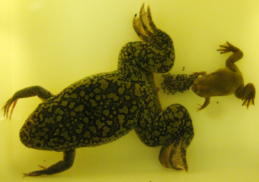
The solution came in the form of a close cousin of Xenopus laevis, Xenopus tropicalis. X tropicalis is smaller than X laevis, has a shorter life cycle, matures in about four months and has a small diploid genome. There were high hopes that X tropicalis would have all the advantages of X laevis and simpler genetics as well.


The genome of Xenopus tropicalis was sequenced in 2010 – the first sequence of an amphibian. The high-quality sequence has aided researcher’s using X tropicalis to have a better understanding of its embryo development and cell biology.
Two Xenopus model organisms
Studying both X laevis and X tropicalis has enabled scientists to discover much about the early three-dimensional development of the embryo after initial fertilisation.
It’s been particularly useful for the analysis of events occurring very early in development. For example, scientists have used X laevis to understand the electrical and nutrient changes that occur as an egg is fertilised, causing the egg to polarise and begin the earliest stages of embryo development.
We’ve also learned more about the development of different systems, including the formation of the neural plate – which gives rise to the entire nervous system. Scientists have also used Xenopus eggs to understand how nerve cells develop in people with different neurological conditions, including autism.
Many questions about development remain unanswered – but without the African clawed frog, our understanding of early development would not have progressed to where it is today.
